Interpreting Echocardiogram Reports
Understanding an Echo Report
Check out this great resource A Practical Approach to Transesophageal Echocardiography
Date of Procedure
This should be within one year of surgery.
Reason for Test
Did the patient status change or is this a normal surveillance echo?
Image Quality
This is mentioned somehwere in the study. Poor quality images can lead to poor conclusions. If this is a TTE study consider TEE for better images.
Rate and Rhythm
Dysrhythmias can make LV function analysis both in systole and diastole anomalus. This can also happen with simple brady or tachycardias.
Chamber Sizes
Check for dilated chambers.
From the pdf:
The quantification of cardiac chamber size and function is the cornerstone of cardiac imaging, with echocardiography being the most commonly used noninvasive modality because of its unique ability to provide real-time images of the beating heart, combined with its availability and portability. Standardization of the methodology used to quantify cardiac chambers is maintained by creating and disseminating official recommendations, which when followed by practitioners provides uniformity and facilitates communication.
Hypertrophy
On a simimlar note check for hypertrophy. Remember LV thickness corresponds to LV mass and a 1mm error can through off the LV mass by 15g or more!
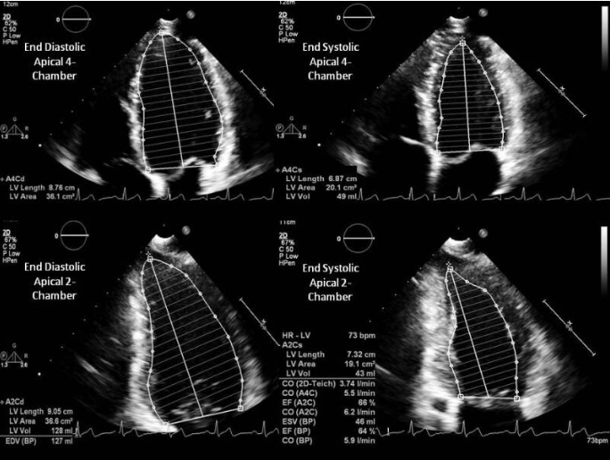
LV Systolic Function
Look for additional notes about systolic function whether its in terms of a percentage or in terms of different parts of the heart are akinetic or hypokinetic. Simnpson’s is the most common method.
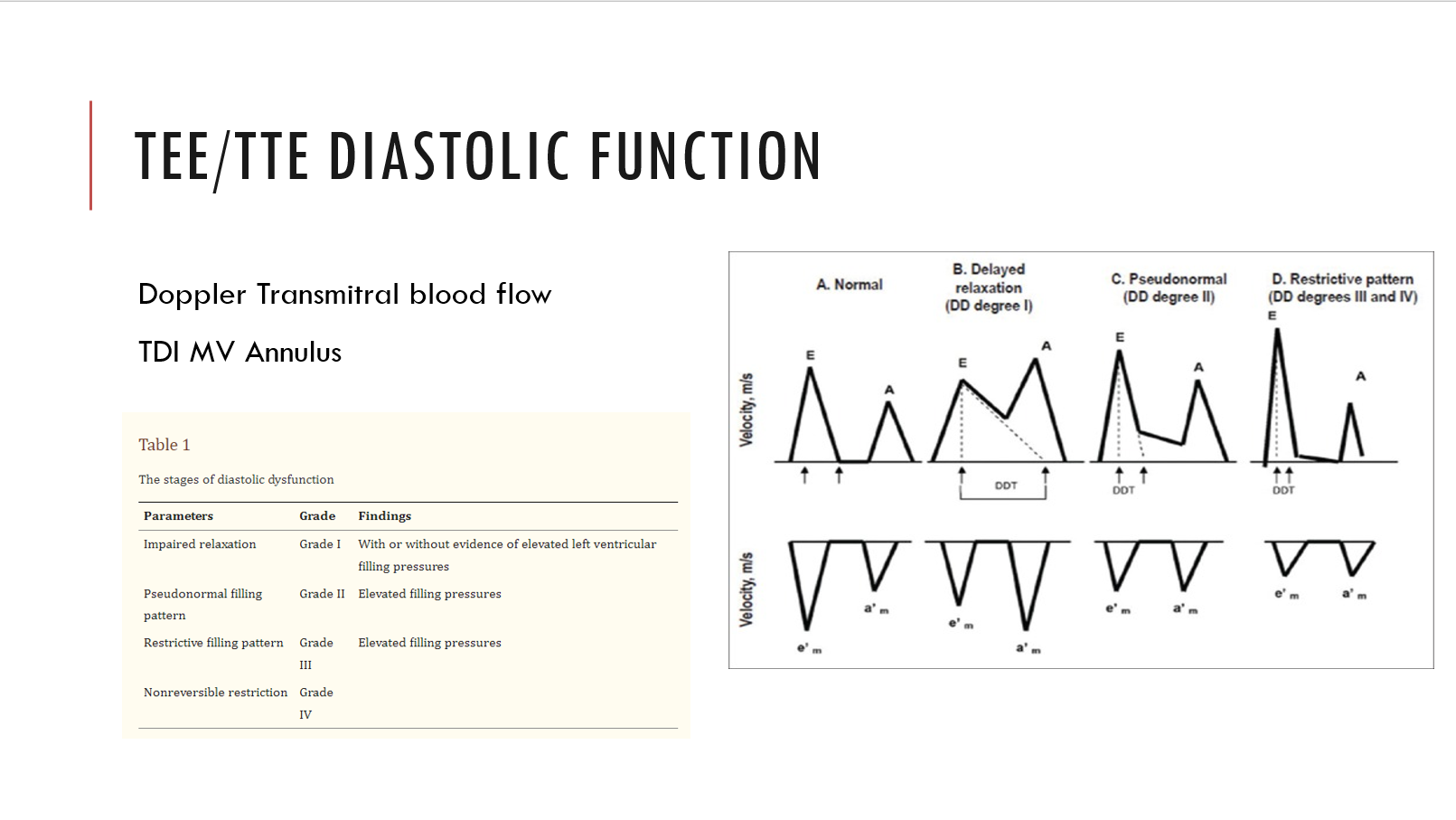
LV Diastolic Function
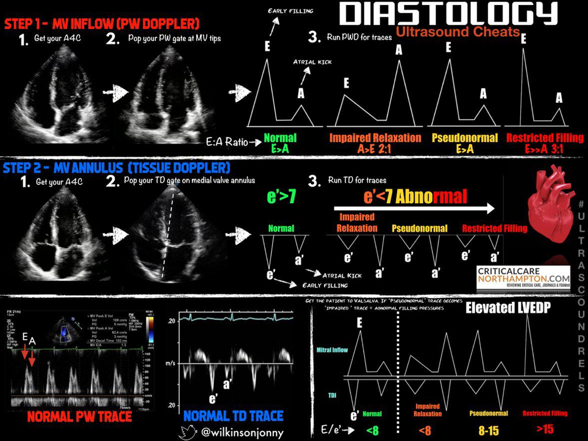
Using Doppler to diagnosis heart failure with preserved ejection fraction requires PW Doppler at the mitral annulus and/or tissue Doppler at the medial mitral annulus. The ratios and velocities are then used to extrapolate diastolic dysfunction.
Grade 1 through 4. Grade 1 is associated with 8x increase in all cause mortality within 5 years!
If e’ is < 7 (MV tissue doppler), or the E/e’ is more than 8, or if the E:A ratio is more than 2:1 there is diastolic dysfunction
Examples of diastology with traditional methods:
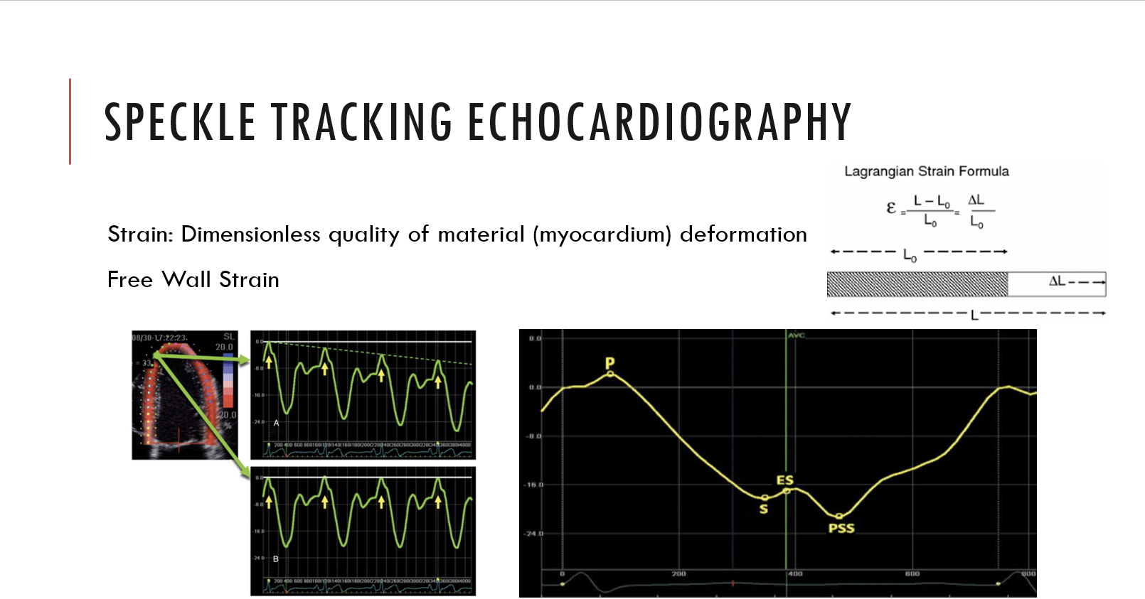
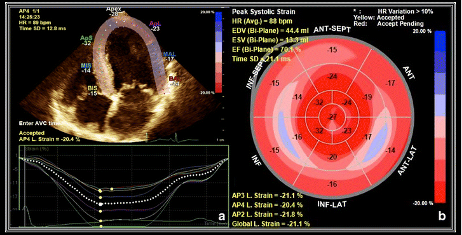
Examples of speckle tracking:
RV
Functional status usually in terms of a percentage.
Valvular Regurgitation and Stenosis
Measurements and grading will be present. Remember a small yet high velocity jet through a small orifice can appear more severe than a larger slower jet in a larger orifice. Use the PISA (proximal isovelocity surface area) quantification of valvular regurgitation to mitigate this effect.
Valve Severity Charts
Mitral Regurgitation
| Measurement | Mild | Moderate | Severe |
|---|---|---|---|
| Vena Contracta (mm) | < 3 | 3-6.9 | > 7 |
| Jet area / LA area | < 20 | 20-40 | > 40 |
| PISA radius (mm) | < 4 | 4-10 | > 10 |
| Regugitant volume (ml) | < 30 | 30-59 | > 60 |
| Regurgitant fraction % | < 30 | 30-49 | > 50 |
| EROA (cm^2) | < 0.2 | 0.2-0.4 | > 0.4 |
Mitral Stenosis
| Measurement | Normal | Mild | Moderate | Severe |
|---|---|---|---|---|
| Valve Area ($cm^2$) | > 2 | 1.5-2 | 1-135 | < 1 |
| Pressure Half Time (ms) | < 90 | 90-150 | > 150 | |
| Indexed Valve Area $cm^2 / m^2$ | 2 | > 0.85 | 0.6-0.85 | < 0.6 |
Aortic Stenosis
| Measurement | Aortic Sclerosis | Mild | Moderate | Severe |
|---|---|---|---|---|
| Valve Area ($cm^2$) | 2.6-3.5 | > 1.5 | 1-1.5 | < 1 |
| Mean Gradient (mmHg) | < 10 | < 20 | 20-40 | > 40 |
| Indexed Valve Area $cm^2 / m^2$ | 2 | > 0.85 | 0.6-0.85 | < 0.6 |
| Peak velocity of flow (m/sec) | 2.6 | < 2.6-3 | 3-4 | > 4 |
Aortic Regurgitation
| Measurement | Mild | Moderate | Severe |
|---|---|---|---|
| Vena Contracta (mm) | < 0.3 | 0.3-0.6 | > 0.6 |
| Jet area / LVOT area | < 4 | 4-6 | > 60 |
| PHT (ms) | > 500 | 500-200 | <200 |
| Regugitant volume (ml) | < 30 | 30-59 | > 60 |
| Regurgitant fraction % | < 30 | 30-49 | > 50 |
| EROA (cm^2) | < 0.1 | 0.1-0.29 | > 0.3 |
Intracardiac Mass
Present or not with sizing and gradients as needed.
Septal Defects
Present or not with flow direction.
RV Systolic Pressure
If RV failure is suspected RVSP are often elevated. This is also elevated in obesity, hypertesnion and pulmonary hypertension.
Pericardium
Effusions if noticed.
Aorta
Calcifications and sizing.
Incidental Findings
Any abnormalities including structural defects, pleural effusions etc.
Summary of Findings
Usually near the beginning or the end and can be useful but be sure to read the corresponding sections for the details.