Interpreting 12 Lead EKGs
EKG Basics
For a great video lecture series about 2.75 hrs long check out EKG interpretation Video Lectures
From the pdf:
Electrocardiography is a fundamental part of cardiovascular assessment. It is an essential tool for investigating cardiac arrhythmias and is also useful in diagnosing cardiac disorders such as myocardial infarction. Familiarity with the wide range of patterns seen in the electrocardiograms of normal subjects and an understanding of the effects of noncardiac disorders on the trace are prerequisites to accurate interpretation.
The Rule of Fours
Four Initial Features
History and Clinical Picture
Clinical picture is arguabley the most important part. History taking is also important but clinical features can be quite informative.
Rate
Normal 60-100. Not too fast or too slow!
Rhythm
Hopefully sinus…but it can be anything! How do you know? First it’s a good idea to know some rhythms and then work through the EKG in an organized manner. You already have the rate so it’s fast, slow, or normal. Next:
- Regular or irregular R-R
- P wave with every QRS
- QRS with every P
- PR interval
- QRS Interval
- Ectopy
- Identify!
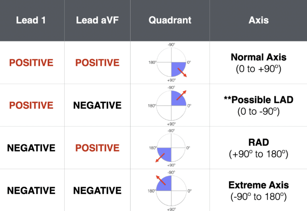
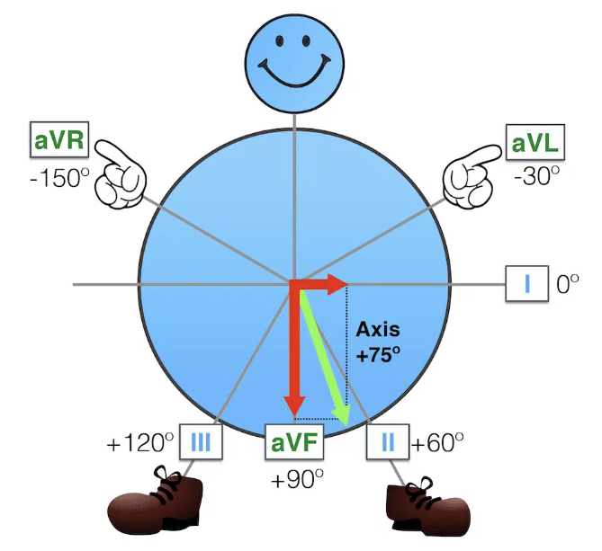
Axis
Normal axis is -30 to + 90.
Axis Deviation In Depth
Check out Life In The Fast Lane for tons of EKG knowledge and examples!
For an in depth analysis of axis deviation check out EKG Axis Interpretation
Ever wonder how the axis is calculated? You use the axis circle and draw vectors. These vectors are summed using the x intercept and the intersection point on the circle. This new vector is the axis deviation! For a thorough explanation and more examples check out Super Axis Man SAM
Four Waves
P
Check out lead II for the best P wave morphology!
QRS
Look in all leads for the presence of Q waves. Also note the QRS amplitude and R wave progression in leads V1-V6.
T
Look in all leads for the presence of T waves. Be sure to note any inverstion, flattening, and the concordance vs discordance with the QURS complex.
U
Are the U waves present or not?
Four Intervals
PR
Normal is 0.12-0.2 seconds or 3-5 little squares. Longer means a first degree heart block, but shorter can be mean WPW, junctional rhythms, and some syndromes.
QRS
Normal is 0.12seconds or less or less than 3 little squares. Widened QRS indicates a conduction defect or delay.
ST
This can be the most important thing at which to look!
QT
Easiest to use the EKG machine measurements and ensure it is not prolonged! ( QT > 450)